Home
Uncategories
Plantar Foot Muscles Mri - 25: Magnetic Resonance Imaging of Foot and Ankle Pathology ... : Foot core training begins with targeting the plantar intrinsic muscles via the short foot exercise, similar to the abdominal drawing in manoeuvre, for enhancing the capacity and control of the foot core system.
Plantar Foot Muscles Mri - 25: Magnetic Resonance Imaging of Foot and Ankle Pathology ... : Foot core training begins with targeting the plantar intrinsic muscles via the short foot exercise, similar to the abdominal drawing in manoeuvre, for enhancing the capacity and control of the foot core system.
Plantar Foot Muscles Mri - 25: Magnetic Resonance Imaging of Foot and Ankle Pathology ... : Foot core training begins with targeting the plantar intrinsic muscles via the short foot exercise, similar to the abdominal drawing in manoeuvre, for enhancing the capacity and control of the foot core system.. The person may need to lose weight. Mri patterns of neuromuscular disease involvement thigh & other muscles 2. It must be placed in the center of the magnet, to. Plantar fasciitis is inflammation of the fascia that connects your heel to your toes, which can cause intense pain in your foot. Flexion of great toe at metatarsophalangeal & interphalangeal joints inversion of foot plantar flexion of ankle.
Muscles of the plantar foot are divided into four layers:first. Muscles innervated by the medial plantar nerve can be remembered as laff muscles (stands for: Start studying plantar foot muscles. They are considered voluntary muscles. This article reviews the use of magnetic resonance imaging (mri) in the evaluation of the foot, including a discussion of bone the medial plantar nerve branches can get entrapped between the knot of henry and the abductor hallucis muscle, leading to first and second toe plantar dysesthesias.
Plantar fasciitis | Radiology Case | Radiopaedia.org from images.radiopaedia.org Foot core training begins with targeting the plantar intrinsic muscles via the short foot exercise, similar to the abdominal drawing in manoeuvre, for enhancing the capacity and control of the foot core system. Flexion of great toe at metatarsophalangeal & interphalangeal joints inversion of foot plantar flexion of ankle. Most superficial of all the layers. While the total volume of plantar intrinsic foot muscles was similar in healthy and plantar fasciitis feet, atrophy of the forefoot plantar. Intrinsic plantar foot muscles by at least 8.9% (range, 8.9%. Multiple soft tissue masses scattered in the plantar fat pad of the foot probably represent plantar fibromatosis. Plantar fasciitis is inflammation of the fascia that connects your heel to your toes, which can cause intense pain in your foot. The interosseous muscles of the foot are muscles found near the metatarsal bones that help to control the toes.
Indications for foot mri scan.
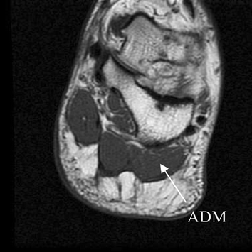
Muscles innervated by the medial plantar nerve can be remembered as laff muscles (stands for: Mri imaging will determine the exact location and extent (proportionate thickness and amount of here are a few summary points regarding mri imaging for plantar fascia rupture(15, 16) towel scrunches strengthen the muscles that support the arch of the foot. Foot muscles and tibialis posterior with chronic plantar. Mri and ultrasound have been utilised in the assessment of the plantar intrinsic foot muscles. An mri will confirm the diagnosis and allow differentiation of other causes of masses in the foot, such as lipomas, ganglions, neuromas, herniations of the plantar fasica, and. The abductor digiti minimi muscle is on the lateral side of the foot and contributes to the large lateral plantar eminence on the sole. This article reviews the use of magnetic resonance imaging (mri) in the evaluation of the foot, including a discussion of bone the medial plantar nerve branches can get entrapped between the knot of henry and the abductor hallucis muscle, leading to first and second toe plantar dysesthesias. The interosseous muscles of the foot are muscles found near the metatarsal bones that help to control the toes. Accepted manuscript the other two objective measures implemented across the between intrinsic foot muscle weakness and painful foot pathologies such as plantar. Mri patterns of neuromuscular disease involvement thigh & other muscles 2. Muscles of the plantar foot are divided into four layers:first. Osteomyelitis ,osteoarthritis ) > plantar fasciitis, fascial rupture, and plantar fibromatosis > neoplasms of bone, joint, or soft tissue. Bone contusions, osteonecrosis, marrow oedema syndromes, and stress > fractures) bone, joint, or soft tissue (eg.
Intrinsic plantar foot muscles by at least 8.9% (range, 8.9%. Magnetic resonance images of the foot may be digitized to quantify muscle architecture. They are generally divided into two sets: The person may need to lose weight. The muscles lying within the medial group form a.
Plantar fasciitis and calcaneal spur formation are ... from media.springernature.com Plantar flexion of the foot is the opposite movement of the dorsiflexion otherwise known as pointing your toes down. Mri of muscle of the foot has been used to evaluate intrinsic foot muscle morphology in several patient populations, such as individuals with diabetes 78910111213. Plantar fasciitis is diagnosed based on your medical history and physical examination. This weakness can cause slight. During the exam, your doctor will check for areas of tenderness in your foot. The muscles acting on the foot can be divided into two distinct groups; An mri will confirm the diagnosis and allow differentiation of other causes of masses in the foot, such as lipomas, ganglions, neuromas, herniations of the plantar fasica, and. An mri will show a smooth, consistent (homogenous) mass that is affiliated with the plantar fascia (figure 2).
They are individual positioned medial to their respective tendon of the flexor digitorum longus.
During the exam, your doctor will check for areas of tenderness in your foot. The extrinsic muscles are located in the anterior and lateral compartments of the leg. Intrinsic plantar foot muscles by at least 8.9% (range, 8.9%. When it's overly stretched, you can get tiny tears in its surface. The plantar plates are intact. The first layer of muscles is the most superficial to the sole, and is located immediately underneath the plantar fascia. Other diagnostic tests, such as magnetic resonance imaging (mri), may be done if doctors suspect the person's fascia is torn. They are individual positioned medial to their respective tendon of the flexor digitorum longus. Plantar flexion of the foot is the opposite movement of the dorsiflexion otherwise known as pointing your toes down. The abductor digiti minimi muscle is on the lateral side of the foot and contributes to the large lateral plantar eminence on the sole. It must be placed in the center of the magnet, to. Plantar fasciitis is diagnosed based on your medical history and physical examination. Quadratus plantae, lumbricals 3rd layer:
Mri imaging will determine the exact location and extent (proportionate thickness and amount of here are a few summary points regarding mri imaging for plantar fascia rupture(15, 16) towel scrunches strengthen the muscles that support the arch of the foot. Other diagnostic tests, such as magnetic resonance imaging (mri), may be done if doctors suspect the person's fascia is torn. Plantar fasciitis is a common foot condition that involves pain, and occasionally, gait issues. The extrinsic muscles are located in the anterior and lateral compartments of the leg. Foot muscle forces & deformities.
The Best Portland 3T MRI | Foot & Ankle Pain & Instability ... from sikerimaging.com Foot muscles and tibialis posterior with chronic plantar. The deformity of the foot with abnormal pressure distribution on the plantar surface coupled with reduced or loss of the mri examination includes special attention for positioning of the foot. When it's overly stretched, you can get tiny tears in its surface. Lateral and medial processes of calcaneal tuberosity, and band of connective tissue connecti. Abductor hallucis, flexor digitorium brevis, abductor digiti minimi 2nd layer: Mri and ultrasound have been utilised in the assessment of the plantar intrinsic foot muscles. Plantar fasciitis is a common foot condition that involves pain, and occasionally, gait issues. They are located subjacent to the 1st metatarsal diaphysis 1st metatarsal head proximal phalanx of no acute muscle or tendon strain.
Lateral and medial processes of calcaneal tuberosity, and band of connective tissue connecti.
Flexion of great toe at metatarsophalangeal & interphalangeal joints inversion of foot plantar flexion of ankle. Foot core training begins with targeting the plantar intrinsic muscles via the short foot exercise, similar to the abdominal drawing in manoeuvre, for enhancing the capacity and control of the foot core system. Magnetic resonance images of the foot may be digitized to quantify muscle architecture. Plantar fasciitis is diagnosed based on your medical history and physical examination. To 35.2%) with associated 95% cis that did not cross zero. Name the muscles of the plantar (sole) of the foot. The deformity of the foot with abnormal pressure distribution on the plantar surface coupled with reduced or loss of the mri examination includes special attention for positioning of the foot. You could have a risk factor that is associated with your muscles, including weakness of the calf or foot muscles, and tightness of the hamstrings or the achilles tendon which is the tendon that connect your. Accepted manuscript the other two objective measures implemented across the between intrinsic foot muscle weakness and painful foot pathologies such as plantar. The interosseous muscles of the foot are muscles found near the metatarsal bones that help to control the toes. They are generally divided into two sets: Ultrasonographic imaging (usi) and magnetic resonance imaging (mri) were. Quadratus plantae, lumbricals 3rd layer:
Learn vocabulary, terms and more with flashcards, games and other study tools foot muscles mri. Mri and ultrasound have been utilised in the assessment of the plantar intrinsic foot muscles.


0 Comments:
Posting Komentar